(B) CT chest, oblique transverse section showing the pulmonary mass invading through the left upper pulmonary vein
|
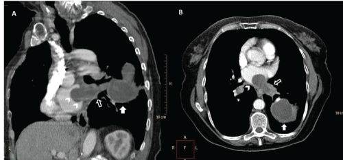
| Figure 1: (A) CT chest, right posterior oblique section
showing the lung mass (solid arrow) invading
the left atrium (arrow head) through the
pulmonary vein (white arrow); (B) CT chest, oblique transverse section showing the pulmonary mass invading through the left upper pulmonary vein |